
ICML-2023 End-to-End Full-Atom Antibody Design
DyMEAN: End-to-end shadow paratope for bridging antibody–antigen design
Abstract
This work introduces dynamic Multi-channel Equivariant grAph Network (dyMEAN), an end-to-end, full-atom antibody design method. The main goal is to improve learning-based antibody design by addressing two central limitations:
- Limitations of prior methods:
- Existing learning approaches often focus on isolated subtasks in the design pipeline, yielding suboptimal overall solutions or excessive resource use.
- Many methods either ignore antibody framework regions or side chains, and thus fail to capture complete full-atom geometric information.
- dyMEAN’s approach:
- End-to-end modeling: dyMEAN directly handles inputs with only an antigen epitope and an incomplete antibody sequence, without a multi-stage pipeline.
- Structural initialization: structure knowledge is used for an initial structural guess.
- Shadow paratope: a shadow paratope bridges antigen–antibody interaction information.
- Adaptive multi-channel equivariant encoder: during full-atom modeling, this encoder adapts to varying atom counts across residues while jointly updating 1D sequence and 3D structure.
- Final docking: antigen–antibody binding structure is predicted by aligning the shadow paratope.
- Experimental validation:
- Experiments on epitope-binding CDR-H3 design, complex structure prediction, and affinity optimization show the benefits of dyMEAN’s end-to-end framework and full-atom modeling.
Introduction
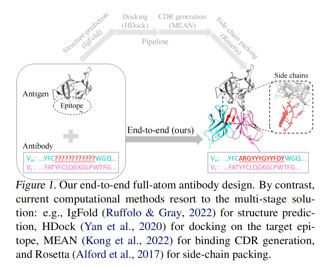
This paper studies antibody design, which is important in therapeutics and biology. Antibody design remains highly challenging because of the high variability of complementarity-determining regions (CDRs) and the complexity of antigen–antibody interaction rules.
Although computational methods have made progress, two core issues persist:
- Many pipelines are multi-stage (structure prediction, docking, CDR generation, side-chain packing), lacking global optimization and leading to suboptimal results or high compute cost.
- Full-atom modeling is often missing, so antigen–antibody complex structures are described imprecisely.
The authors propose dyMEAN (dynamic Multi-channel Equivariant grAph Network), an end-to-end full-atom antibody design model, to address these issues. From an antigen epitope and an incomplete antibody sequence, the model generates a complete 1D sequence and 3D structure, and improves design accuracy and docking via a shadow paratope mechanism and an E(3)-equivariant encoder.
Limitations of existing computational approaches
- Energy-based methods (e.g., Rosetta) optimize antibody structures with statistical energy functions, but expressiveness is limited and antigen–antibody interactions are hard to model accurately.
- Language models (LMs) trained on antibody 1D sequence alone lack 3D geometry, which restricts sequence generation quality.
- Deep generative joint sequence–structure design can generate CDR sequence and 3D structure together, but still suffers from incomplete contextual modeling.
To overcome these issues, dyMEAN is designed with the following properties:
-
It generates the full antibody sequence and 3D structure directly from an antigen epitope and incomplete antibody sequence, without a multi-stage pipeline, reducing error accumulation and compute.
-
It builds an initial structure for framework regions using conserved residues.
-
A shadow paratope is created near the epitope such that it:
- Shares the hidden states of the true CDR-H3.
- Optimizes coordinates independently, so antibody–antigen interactions do not depend on the initial antibody pose.
-
Adaptive multi-channel equivariant encoder
-
Uses E(3)-equivariant updates for 1D sequence and 3D structure jointly.
-
Handles variable atom counts per residue for full-atom modeling.
-
-
Docking is performed by aligning the real CDR-H3 to the shadow paratope, yielding the final antigen–antibody complex.
Notations and Definitions
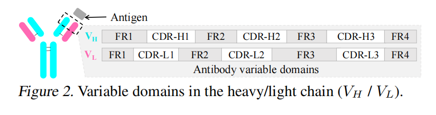
An antibody is a Y-shaped symmetric protein built from two identical chains, each comprising:
- Heavy chain (VH)
- Light chain (VL)
Each chain contains multiple constant domains and one variable domain:
- Constant domains are conserved across antibodies.
- Variable domains mediate antigen-specific binding and are the main focus of design.
We denote the variable domains of the heavy (VH) and light (VL) chains as $V_H$ and $V_L$. Each variable domain alternates four framework regions (FRs) and three complementarity-determining regions (CDRs).
The antibody binding site is the paratope; the antigen binding site is the epitope. Here, paratope refers specifically to CDR-H3, which dominates antigen binding.
-
Graph representations of antibody and antigen
- Epitope graph:
$G_E(V_E,E_E)$ - Antibody graph:
$G_A(V_A,E_A)$
- Epitope graph:
-
Residue features
- Amino acid type
$s_i$: the residue identity (e.g., Ala, Ser, Glu). - Multi-channel 3D coordinate matrix
$X_i\in \mathbb{R}^{3\times c_i}$$c_i$is the number of atoms in the residue (backbone and side chain).- Because side chains differ,
$c_i$is not fixed across amino acids. - dyMEAN uses a multi-channel representation to handle variable atom counts.
- Amino acid type
-
Graph edge construction
-
dyMEAN builds edges with k-nearest neighbors (kNN) using the minimum pairwise atom distance between residues
$v_i$and$v_j$:
where p and q index atom coordinates.
-
-
Three global nodes are added on the heavy chain, light chain, and antigen epitope.
Task Definition
Given antigen epitope + partial antibody sequence, predict the full CDR-H3 sequence and 3D structure such that the result docks correctly to the epitope.
Our Method: dyMEAN
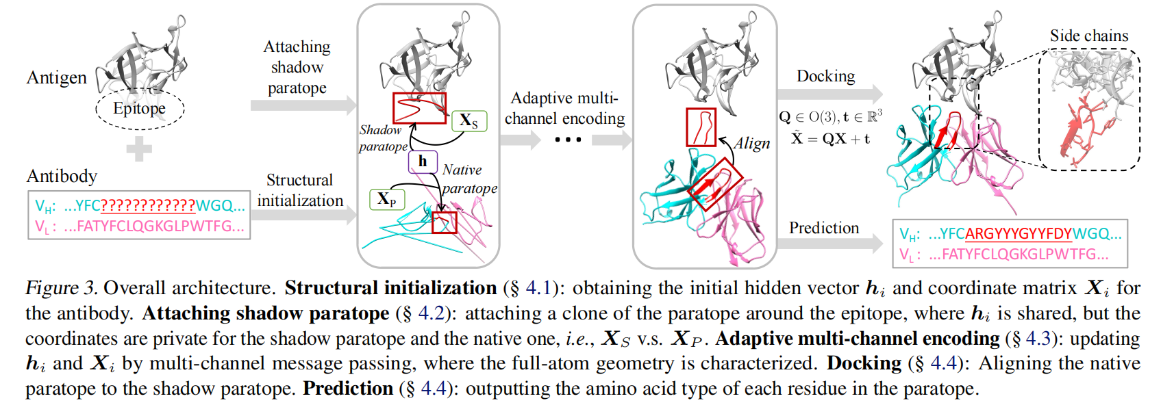
Each vertex (from epitope graph $G_E$, antibody graph $G_A$, and paratope subgraph $G_P$) carries:
- An invariant vector
$h_i\in\mathbb{R}^d$ - An equivariant coordinate matrix
$X_i\in\mathbb{R}^{3\times c_i}$
The overall pipeline is:
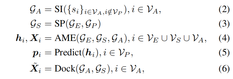
where:
- SI (Structural Initialization):
- From the known incomplete antibody sequence, predict initial coordinates
$X_i^{(0)}$and hidden states$h_i^{(0)}$.
- From the known incomplete antibody sequence, predict initial coordinates
- SP (Shadow Paratope):
- Insert a shadow paratope
$G_S$between epitope and paratope; it shares CDR-H3 hidden states, connects antigen and antibody, and anchors docking.
- Insert a shadow paratope
- AME (Adaptive Multi-channel Encoder):
- Iteratively update hidden states
$h_i$and coordinates$X_i$for all vertices.
- Iteratively update hidden states
- Prediction:
- Compute the CDR-H3 amino acid distribution
$p_i$.
- Compute the CDR-H3 amino acid distribution
- Docking:
- Complete antibody–antigen binding by aligning the shadow paratope
$G_S$.
- Complete antibody–antigen binding by aligning the shadow paratope
Structural Initialization with Conserved Residues
In the input, antibody sequence $\{s_i\}_{i\in V_A,i\notin V_P}$ lacks CDR-H3 and 3D structure is unknown. dyMEAN must initialize antibody hidden states $h_i^{(0)}$ and 3D coordinates $X_i^{(0)}$.
Hidden state initialization $h_i^{(0)}$
Each residue hidden state combines amino acid type and position encoding: $h_i^{(0)}=f(s_i,r_i)=f_{s_i}+f_{r_i}$. Missing CDR positions use a special [MASK] token.
3D coordinate initialization $X_i^{(0)}$
Framework regions (FRs) are typically conserved. dyMEAN initializes 3D coordinates ($X_i^{(0)}$) from framework coordinates in known antibody structures as follows:
- Align conserved residues:
- Compare the input antibody sequence to sequences in a structure database and identify residues conserved across many antibodies.
- Antibody numbering (e.g., IMGT) is used to find residues with consistent type in more than 95% of antibodies.
- Kabsch alignment:
- Kabsch algorithm aligns backbone atoms of conserved framework residues to obtain average 3D coordinates for initialization.
- Interpolation and extension:
- Non-conserved framework residues get coordinates by linear interpolation between conserved ones; terminal residues use outward linear extrapolation from the nearest conserved residue.
Coordinate normalization
After initialization, dyMEAN normalizes coordinates:
- 3D translation so coordinates have zero mean.
- Per-dimension variance scaling for consistent scale across antibodies.
This yields a reasonable initial guess for framework regions, supporting subsequent shadow paratope construction and multi-channel message passing.
E(3)-Invariant Attachment of Shadow Paratope
To strengthen antigen–antibody information exchange, dyMEAN attaches a Shadow Paratope near the epitope—a pseudo paratope copy with two roles:
- E(3)-invariant information transfer: the shadow paratope shares hidden states
$h_i$and topology with the true paratope, enabling exchange without relying on the antibody’s initial pose. - Docking anchor: the shadow paratope is central to docking (see §4.4).
Because only E(3)-invariant information (hidden states $h_i$) is shared—not coordinates $X_i$—3D coordinates and final docked structures are insensitive to initial antibody placement, improving robustness.
Constructing the shadow paratope
The shadow paratope is a subgraph $G_s=(V_S,E_S)$ where:
$V_S$contains shadow paratope residues.$E_S$has two parts:- Internal edges: copied from the original paratope.
- External edges: connect shadow paratope to the epitope.
For epitope residue $v_i \in V_E$ and shadow paratope vertex $v_j \in V_S$, external edges use kNN distance:

where $\varphi_e$ is an MLP (Multi-Layer Perceptron) that learns distance features.
Initializing the shadow paratope
- Hidden vectors
$h_i$are copied from the true paratope. - Coordinates
$X_i$are drawn from a standard Gaussian$N(0, I)$near the epitope center, adding early-training randomness without breaking E(3) invariance.
Finally, $G_S$ is merged into $G_E$ as $G_E \cup G_S$ for message passing and docking.
Adaptive Multi-Channel Equivariant Encoder
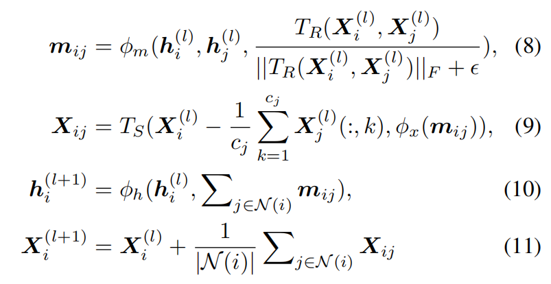
Geometric Relation Extractor $T_R$
-
Channel-wise Euclidean distances: for
$X_i\in\mathbb{R}^{3\times c_i}$and$X_j\in\mathbb{R}^{3\times c_j}$, each channel (atom) uses$D_{ij}(p, q) = ||X_i(:, p) - X_j(:, q)||_2$. -
Weighted correlation:

with
$w_{i}\in\mathbb{R}^{c_i\times 1}$,$w_{j}\in\mathbb{R}^{c_j\times 1}$,$A_{i}\in\mathbb{R}^{c_i\times d}$, and$A_{j}\in\mathbb{R}^{c_j\times d}$.The output
$R_{i,j}\in\mathbb{R}^{d\times d}$has fixed dimension for consistent inputs.
Geometric Message Scaler, $T_S$
$T_S$ scales geometric information so coordinates from different residues fuse effectively. For coordinates $X\in\mathbb{R}^{3\times c}$ and non-geometric $s=\phi_x(m_{ij})\in\mathbb{R}^C$ (C is an upper bound on channel count), $T_S(X,s)$ is:

where $s'\in\mathbb{R}^c$ is mean-pooled s, window size C−c+1, stride 1, and diag(·) takes matrix diagonals.
Information exchange between $G_E$ and $G_A$
Epitope graph $G_E$ and antibody graph $G_A$ are not directly connected initially, yet the model must exchange information for antibody–antigen docking. dyMEAN routes messages through the shadow paratope ($G_S$).
- Stage 1 (Antibody graph
$G_A$)- Run one layer of AME on
$G_A$. - Copy hidden vectors
$h_i$from paratope$G_P$to shadow paratope$G_S$, so$G_S$carries paratope information.
- Run one layer of AME on
- Stage 2 (Epitope graph
$G_E$and shadow paratope$G_S$)- Run one layer of AME on
$G_E \cup G_S$to exchange epitope and shadow paratope information. - Copy hidden vectors from
$G_S$back to paratope$G_P$, injecting epitope context into the antibody.
- Run one layer of AME on
- Alternating iterations
- Repeat the two stages for L layers, gradually converging cross-molecule information.
- Then run one extra AME layer on
$G_A$to propagate updates through the full antibody.
The Geometric Relation Extractor (TR) and Geometric Message Scaler (TS) satisfy:
- TR is
$E(3)$-invariant: geometric relations are unchanged under rotation, translation, and reflection. - TS is
$O(3)$-equivariant: message passing respects rotations. - Exchange
$G_E ↔ G_A$via$G_S$is$E(3)$-invariant.
Prediction, Docking and Training Losses
Prediction
dyMEAN uses progressive full-shot decoding to predict CDR-H3 1D sequence and 3D structure, refined over T iterations:

An MLP predicts CDR-H3 amino acids:

Hidden states $h_i^{(t)}$ update each round:

where $f(s_i,r_i)=f_{s_i}+f_{r_i}$ embeds sequence position, $p_i^{(t)}(j)$ is the probability of amino acid j at CDR-H3 position i, and $\phi_d(h_i^{(t)})$ is a memory term.
After each iteration, dyMEAN recomputes edges between $G_A$ and $G_E$ to refresh topology.
Docking
After the final iteration, dyMEAN docks with the Kabsch algorithm (Kabsch, 1976):

-
Shadow paratope
$G_S$provides a reference pose for predicted CDR-H3. -
Kabsch alignment moves CDR-H3 (
$V_P$) onto shadow paratope ($V_S$) to optimize the complex.
Loss function
Cross-entropy supervises CDR-H3 sequence prediction:

Huber loss trains final 3D coordinates:

A chemical bond-length term is added:

Total structure loss is the sum of coordinate and bond terms.
For docking quality, dyMEAN adds shadow paratope coordinate loss and external distance loss:
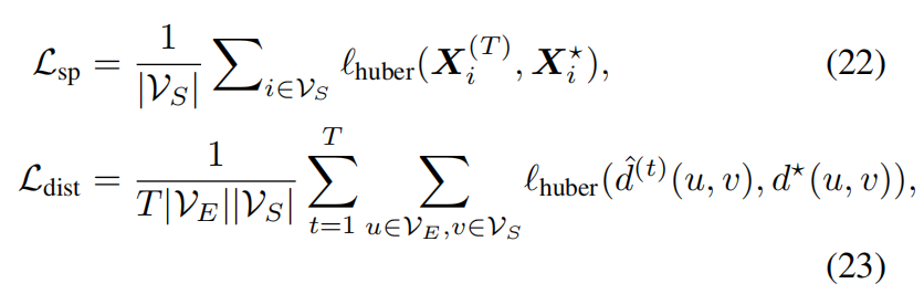
Final docking loss is the sum of both.
Experiments
dyMEAN is trained on SAbDab with PyTorch distributed training.
2.1. Training hyperparameters
Training details
- Optimizer: Adam
- Initial learning rate:
$1 \times 10^{-3}$, exponential decay to$1 \times 10^{-4}$. - Batch size: 16
- Epochs:
- CDR-H3 design & affinity optimization: 200
- Complex structure prediction: 250
- Learning rate: cosine schedule to reduce overfitting.
Masking
Because CDR-H3 is the main variable region, training uses dynamic masking:
- Early training: 90% of paratope residues remain unmasked.
- Later training: masking increases until all paratope residues are masked, so the model can generate sequences under missing residues.
Dataset and baselines
-
The 48 epitope residues closest to the antibody are extracted, sufficient to cover binding residues.
-
Because prior end-to-end full-atom antibody design methods were scarce, the authors compare to:
- IgFold (Ruffolo & Gray, 2022): AlphaFold-style antibody structure prediction.
- HDock (Yan et al., 2020): knowledge-based docking scores.
- RosettaAb (Adolf-Bryfogle et al., 2018): statistical energy optimization of sequence and structure.
- MEAN (Kong et al., 2022): equivariant attention graph networks for CDR-H3 generation.
- Diffab (Luo et al., 2022): diffusion-based CDR generation with side-chain orientation.
- HERN (Jin et al., 2022): end-to-end design without separate structure prediction, docking, or side-chain packing, but without framework modeling.
Epitope-binding CDR-H3 Generation
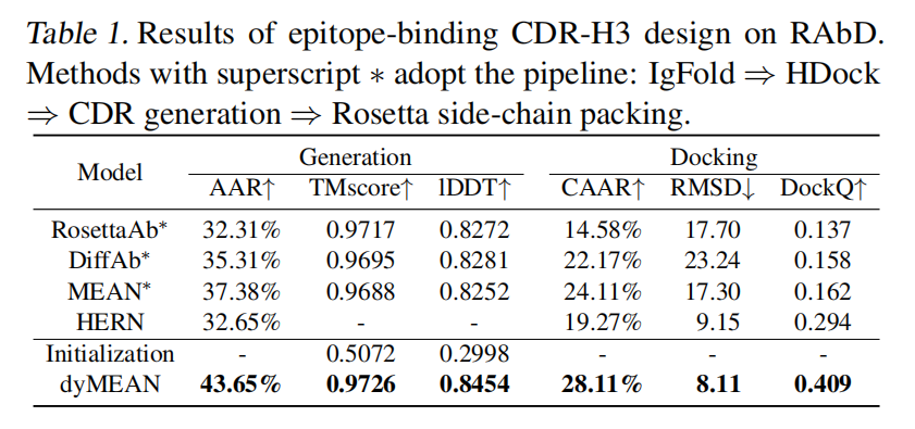
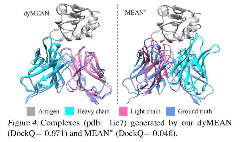
Goal: CDR-H3 is the most variable region and sets binding specificity and affinity (Raybould et al., 2019). The task is to predict CDR-H3 1D sequence and 3D structure.
Metrics
- Amino Acid Recovery (AAR): overlap between generated and native sequence.
- Contact AAR (CAAR): AAR for CDR-H3 residues within 6.6Å of the epitope (Ramaraj et al., 2012).
- TM-score: global similarity of predicted vs. native CDR-H3 structure (Zhang & Skolnick, 2004).
- Local Distance Difference Test (lDDT): similarity from inter-atomic distance matrices (Mariani et al., 2013).
- RMSD: root-mean-square deviation of CDR-H3 coordinates without Kabsch alignment.
- DockQ: composite docking quality (Basu & Wallner, 2016).
Results
Table 1 (CDR-H3 design): dyMEAN achieves AAR = 43.65%, TM-score = 0.9726, lDDT = 0.8454, DockQ = 0.409, outperforming baselines on all reported metrics.
Complex Structure Prediction
Goal: given CDR-H3 sequence, predict the 3D structure of the full antibody–antigen complex.
Comparison
Baselines include:
- IgFold⇒HDock (IgFold backbone, then HDock)
- IgFold⇒HERN (HERN docking, Rosetta side chains)
- GT⇒HERN (HERN with ground-truth structure)
- dyMEAN (end-to-end full-atom prediction)
Results (Table 2)
dyMEAN exceeds baselines on all metrics.
Even when HERN uses ground-truth antibody structure (GT⇒HERN), dyMEAN remains stronger, suggesting effective modeling of antibody–antigen interactions.
Affinity Optimization
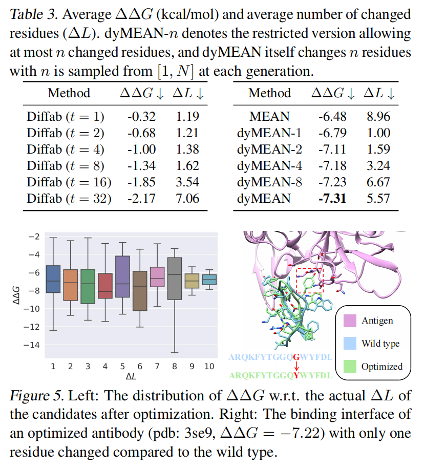
Goal: optimize sequence to maximize binding affinity, measured by ΔΔG (lower is better binding).
Protocol
- GNN-based ΔΔG predictor (Shan et al., 2022).
- FoldX as an affinity scoring tool.
- ΔL (number of mutated residues) to avoid excessive mutation.
Results (Table 3)
dyMEAN achieves larger ΔΔG improvements than MEAN and DiffAb with lower ΔL.
Compared to MEAN (ΔΔG = −5.84, ΔL = 5.09) and DiffAb, dyMEAN shows stronger affinity optimization.
Analysis
Ablation study
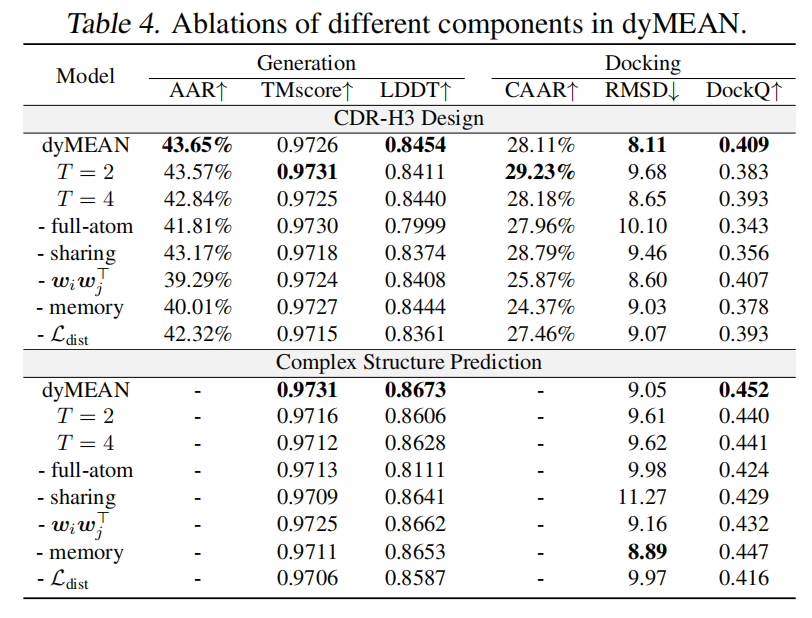
Ablations quantify component contributions.
Results (Table 4)
- T mainly affects docking; T = 3 is best.
- Removing full-atom modeling sharply hurts performance—side-chain conformations matter.
- Removing shadow paratope sharing drops most metrics (except CAAR), showing its importance for structure and docking.
- Removing learnable channel weights degrades performance—channel weights act like attention over atoms.
- Memory helps CDR-H3 design more than complex prediction: sequence hidden states use memory strongly; 3D coordinates propagate directly.
$L_{dist}$is critical for docking; coordinates alone may not recover shadow paratope structure early in decoding.
Multiple CDRs design and full antibody design
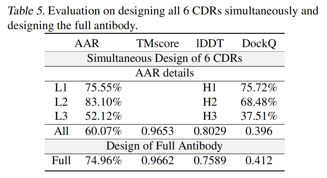
Multiple CDRs design: generate with CDR-H1, H2, H3 and L1, L2, L3 all masked.
Full antibody design: generate the entire antibody including framework regions (FRs).
Limitations
Data diversity and evaluation metrics
Deep generative antibody design still struggles partly because antigen–antibody data lack diversity.
Analyzing the most frequent single-letter patterns at each CDR-H3 position (matched from ends toward the center) reveals a dominant motif “ARDG***DY”, with many “*” positions as Y.
-
Applying this unigram pattern on the test set yields AAR = 39.61%, CAAR = 26.57%.
-
Meaningless unigram patterns dominate train and test, which may limit learning of true antigen–antibody interactions.
-
After removing the first 4 and last 2 CDR-H3 residues, dyMEAN’s AAR drops to 31.76%, showing frequent unigrams inflate sequence metrics.
Implications:
- Datasets may need enrichment (lab data, or interfaces from broader protein complexes).
- Metrics may need refinement, e.g.:
- Exclude residues predictable from unigrams to reduce bias.
- Use richer criteria for plausible antigen–antibody interactions.
Reliability of computational energy functions
Ultimately, binding affinity determines whether designed candidates are useful. This work uses a deep learning ΔΔG predictor. Computational energy methods remain uncertain:
Statistical energy tools include FoldX (Schymkowitz et al., 2005), Rosetta (Alford et al., 2017), and docking score functions (Goodsell et al., 1996).
Issues
- Reliability is still uncertain; several correlate poorly with experiment (Ramírez & Caballero, 2016, 2018).
- Open questions:
- Can these scores separate weak binders?
- Do functions trained on native complexes generalize to deep-learning-generated structures with different statistics?
Conclusion
- Without reliable computational affinity scores, wet-lab validation remains necessary, at higher cost and time.
- Future work should build more generalizable affinity predictors to improve reliability of learning-based antibody design.